Point‑of‑Care Ultrasound Training: A Practical Guide to POCUS Case Libraries, Reference Guides, and Learning Modules
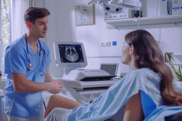
Point‑of‑care ultrasound (POCUS) has moved from a niche skill to a core part of everyday clinical practice in many healthcare settings. Whether you work in emergency medicine, critical care, internal medicine, primary care, anesthesia, or another field, POCUS can change how you assess patients at the bedside.
Yet one of the biggest challenges for clinicians is how to train effectively:
- Where do you find high‑quality POCUS case libraries?
- Which reference guides are actually practical when you are on shift?
- How do you structure your learning with modules and curricula instead of random videos and one‑off workshops?
This guide walks through the main types of resources available, how to access and use them strategically, and how to build a sustainable POCUS learning pathway without getting overwhelmed.
Why POCUS Training Needs More Than a Single Course
Many clinicians are first exposed to POCUS in a brief in‑person workshop, a residency rotation, or a short course. These can be helpful for basic familiarity, but they rarely provide enough:
- Image interpretation skills grow through repeated exposure to varied cases.
- Probe handling and scanning technique improve through deliberate, focused practice.
- Clinical integration develops when you see how POCUS findings change decisions and outcomes over time.
Because of this, many educators emphasize a combination of:
- Structured learning modules (for core theory and image patterns)
- Case libraries (for pattern recognition and clinical reasoning)
- Quick‑access reference tools (for use at the bedside and on call)
Understanding how these pieces fit together helps you spend your limited time more efficiently.
Mapping Out Your POCUS Learning Journey
Before diving into specific resource types, it helps to frame POCUS training in stages. These stages often overlap, but each benefits from different tools.
H2: Common Stages of POCUS Skill Development
H3: 1. Orientation: Getting Familiar with the Basics
At this stage, the goals are:
- Understanding machine controls (gain, depth, presets, probe types)
- Knowing basic safety and limitations
- Learning standard views for core applications (e.g., cardiac, lung, abdominal, vascular)
Useful resources at this stage:
- Introductory learning modules with diagrams and short videos
- Basic reference guides on knobology and anatomy
- A small selection of classic cases showing normal findings
H3: 2. Skill Building: Repetition and Pattern Recognition
Once you can obtain basic images, the emphasis shifts to:
- Distinguishing normal vs. abnormal
- Recognizing common pathologies
- Improving speed and consistency in image acquisition
Useful resources:
- Case libraries with labeled images and clips
- Modular courses focusing on specific systems (lung, cardiac, abdominal, DVT)
- Structured scanning checklists and bedside reference tools
H3: 3. Clinical Integration: Applying POCUS in Real‑World Contexts
Here, you are focusing on:
- Using POCUS as part of a broader clinical assessment
- Understanding indications and pitfalls
- Communicating findings and documenting appropriately
Useful resources:
- Cases with full clinical context (history, exam, labs, imaging follow‑up)
- Advanced reference guides (differential patterns, algorithms)
- Modules on documentation, billing, and quality assurance, where relevant
H3: 4. Refinement: Advanced Views and Teaching Skills
For more experienced users, goals may include:
- More complex applications (advanced cardiac, advanced hemodynamics, MSK, nerve blocks)
- Quality improvement and protocol development
- Coaching or teaching other clinicians
Useful resources:
- Specialized advanced case libraries
- Guideline‑based reference content
- Educator‑oriented teaching modules and assessment tools
Understanding POCUS Case Libraries
POCUS case libraries are collections of real or simulated ultrasound cases, often including:
- Image clips and stills
- Brief clinical summaries
- Key findings and interpretations
- Sometimes follow‑up imaging or outcomes
They can be text‑based, video‑based, or interactive.
H2: Why Case Libraries Matter for POCUS Training
Pattern recognition is central to ultrasound. Seeing many variations of “normal” and “abnormal” across different body types, ages, and disease processes helps clinicians:
- Identify subtle pathology
- Recognize common artifacts
- Avoid over‑calling normal variants
- Understand how POCUS correlates with clinical findings and other imaging
Case libraries also support:
- Independent study between shifts
- Flipped classroom models (reviewing cases before in‑person sessions)
- Group teaching sessions and journal clubs
H2: Types of POCUS Case Libraries
H3: 1. Specialty‑Specific Libraries
Some collections focus on particular fields, such as:
- Emergency and critical care: Shock, cardiac arrest, trauma, respiratory failure
- Internal medicine and hospital medicine: Volume assessment, heart failure, ascites, pleural effusions
- Anesthesia and perioperative care: Nerve blocks, gastric assessment, cardiac function
- Primary care and outpatient settings: Obstetric scanning within scope, musculoskeletal complaints, basic abdominal views
These are particularly useful if you primarily practice in one environment and want highly contextual cases.
H3: 2. Application‑Specific Libraries
Others are organized by ultrasound application:
- Cardiac POCUS (LV function, RV strain, pericardial effusion)
- Lung ultrasound (pneumothorax, consolidation, edema, interstitial syndrome)
- Abdominal and FAST (free fluid, AAA, hydronephrosis)
- Vascular (DVT assessment, line guidance, IVC)
- Soft tissue and musculoskeletal (abscess vs. cellulitis, joint effusions)
This format is ideal when you want to master one application at a time.
H3: 3. Mixed‑Level Collections
Some libraries contain a blend of basic and advanced cases, with:
- Simple “spot the effusion” examples
- More complex multi‑system cases requiring integration of findings
- Pitfalls and mimics (e.g., lung consolidation vs. atelectasis, artifact vs. true pathology)
These collections can be revisited over months or years as your skills grow.
How to Use POCUS Case Libraries Effectively
Having a library is one thing; using it wisely is another. Many learners get more value by following a structured approach rather than passively watching clips.
H2: A Simple Method to Maximize Learning From Case Libraries
🧠 Step‑by‑step approach
Hide the diagnosis first
- Start with the clinical vignette (if available) and the ultrasound clip without reading the answer.
- Ask yourself: What question is the ultrasound trying to answer?
Describe, don’t label
- Verbally or in writing, describe what you see:
- “There is an anechoic fluid collection around the heart,”
- rather than immediately saying, “This is tamponade.”
- This strengthens observation skills, which are essential for accurate interpretation.
- Verbally or in writing, describe what you see:
Commit to an interpretation
- Decide: normal, abnormal, unclear, or not adequately visualized.
- If abnormal, note possible differentials.
Compare with the official explanation
- Read the provided interpretation and teaching points.
- Note differences between your thought process and the case solution.
Extract 1–2 key takeaways per case
- For example:
- “Pleural effusion often layers dependently and may show spine sign.”
- “Large pericardial effusions can still have preserved LV function on basic views.”
- Write these in a notebook or digital note system.
- For example:
Reinforce over time
- Revisit challenging cases weeks later.
- Spaced repetition helps consolidate pattern recognition.
Quick Cheat‑Sheet: Getting the Most from Case Libraries 📚
- 🔍 Focus on description before diagnosis
- 📝 Keep a running log of image patterns and pearls
- ⏱️ Short, regular sessions (10–20 minutes) often work better than rare, long study blocks
- 👥 Discuss selected cases with colleagues or mentors when possible
- 🔄 Revisit tricky cases periodically to see your progress
POCUS Reference Guides: Your On‑Shift Safety Net
While case libraries build your pattern recognition, reference guides help you:
- Remember probe positioning and standard views
- Recall measurement cutoffs and general normal ranges
- Walk through simple decision pathways (for example, approaches to undifferentiated shock)
- Quickly review artifacts and pitfalls
They are not meant to replace formal training or clinical judgment, but they can make your practice more consistent and structured.
H2: Types of POCUS Reference Tools
H3: 1. Pocket Cards and Laminated Sheets
These often cover:
- Basic cardiac, lung, abdominal, and vascular views
- Probe markers and orientation
- Suggested scanning sequences (e.g., FAST exam steps)
They are popular because they are:
- Portable (fit in a pocket or on a workstation)
- Durable (laminated, easy to wipe down)
- Simple (few words, more diagrams)
H3: 2. Digital Reference Apps and Tools
Mobile or tablet‑based references may provide:
- Static images and short clips demonstrating normal and abnormal findings
- Step‑by‑step scanning protocols
- Basic measurement tips and common interpretation frameworks
Many clinicians find digital references convenient for:
- Reviewing just before performing an exam
- Refreshing knowledge during quieter moments on shift
H3: 3. Institution‑Specific Protocol Guides
Hospitals or training programs may create their own:
- POCUS protocols aligned with local practice patterns
- Documentation and billing templates
- Quality assurance checklists
These are especially important because they:
- Reflect what is expected in your own workplace
- Help align your practice with local standards and processes
How to Integrate Reference Guides Into Everyday Practice
To move from theory to habit, many clinicians:
- Keep 1–2 favorite references only, rather than juggling many; this reduces decision fatigue.
- Use the same protocol every time for a given exam (e.g., always scan lung fields in the same order).
- Review reference content briefly before or immediately after performing a scan, to close feedback loops.
- Collaborate with colleagues to standardize reference tools across a team or department.
Over time, the reference becomes less of a crutch and more of a safety net—there when you need to double‑check a detail or confirm a measurement.
Learning Modules: Building a Structured POCUS Curriculum
Case libraries and reference cards are powerful, but they work best when anchored by structured learning modules. These modules provide:
- A clear sequence from basic to intermediate to advanced topics
- Objectives and outcomes for each section
- Content that blends theory, images, and clinical context
H2: What Makes a Good POCUS Learning Module?
Well‑designed modules often have:
- Clear goals (for example, “By the end, you can obtain standard parasternal and apical cardiac views and identify gross LV dysfunction.”)
- Short, focused sections (e.g., 10–20 minutes each)
- A mix of text, images, and video clips
- Interactive elements, such as quizzes or self‑checks
- Opportunities to apply knowledge through cases or practice tasks
H2: Common POCUS Module Categories
H3: 1. Foundational Modules
Typical topics:
- Ultrasound physics basics (kept practical and brief)
- Machine controls and settings
- General scanning principles and ergonomics
- Sterility and infection prevention for procedures
These modules help you understand why images look the way they do and how to optimize them.
H3: 2. Core Clinical Applications
Frequently covered areas:
- Cardiac POCUS: Basic views, global function, pericardial effusion
- Lung and pleural: A‑lines, B‑lines, consolidations, effusions, pneumothorax patterns
- Abdominal and FAST: Free fluid, bladder, basic biliary, aorta screening
- Vascular access and DVT scanning: Vein identification, compression techniques
Modules in this category help clinicians perform the most commonly used bedside studies in acute and general settings.
H3: 3. Procedure‑Guidance Modules
For clinicians whose scope includes procedures, modules may cover:
- Ultrasound‑guided central or peripheral venous access
- Thoracentesis and paracentesis
- Nerve blocks and regional anesthesia
- Abscess drainage guidance
The emphasis tends to be on:
- Needle visualization
- Avoiding nearby structures
- Simple steps to improve safety
H3: 4. Advanced and Specialized Modules
Examples include:
- Advanced cardiac assessment (valve pathology, diastology for selected users)
- Hemodynamic assessment and complex shock states
- Advanced lung and diaphragm evaluation
- Focused obstetric applications appropriate to scope and training
- Musculoskeletal and nerve evaluation
These are generally most helpful once foundational skills are solid.
Combining Case Libraries, Reference Guides, and Modules Into a Cohesive Plan
Instead of using each type of resource in isolation, many learners get better results by intentionally combining them.
H2: A Sample 3‑Month POCUS Learning Framework
Below is a conceptual example of how someone might structure a learning period. It can be adapted based on specialty, schedule, and experience level.
| Timeframe | Primary Focus | Role of Case Libraries | Role of Reference Guides | Role of Learning Modules |
|---|---|---|---|---|
| Weeks 1–2 | Orientation | Review a few classic “normal” cases for each core view | Use basic knobology and view cards during every scan | Complete foundational modules on physics, machines, and basic anatomy |
| Weeks 3–6 | Skill Building | Work through 2–3 cases per week in one system (e.g., lung) | Use standard scanning protocols at the bedside | Complete core application modules (lung, cardiac, abdominal) |
| Weeks 7–10 | Integration | Choose multi‑system cases; focus on decision‑making | Rely on reference guides only as needed; start internalizing steps | Explore modules on documentation, common pitfalls, and QA |
| Weeks 11–12 | Refinement | Revisit earlier challenging cases to gauge progress | Consult advanced references for select scenarios | Begin advanced or specialized modules as appropriate |
This table is simply a planning template, not a requirement. The key concept is to:
- Learn core theory (modules)
- Practice pattern recognition (cases)
- Support real‑time practice (reference tools)
all in parallel.
Adapting POCUS Training to Different Clinical Roles
POCUS training is not one‑size‑fits‑all. The ideal mix of case libraries, references, and modules depends heavily on clinical role and scope.
H2: Common User Profiles and Training Priorities
H3: Emergency and Critical Care Clinicians
Training priorities often include:
- Rapid assessment of shock and respiratory failure
- Resuscitation‑focused cardiac and lung POCUS
- Abdominal trauma and FAST exams
- Vascular access and procedural guidance
Useful emphasis:
- Case libraries with unstable patients and time‑sensitive decisions
- Reference tools for shock and dyspnea algorithms
- Modules on integration with resuscitation protocols
H3: Hospitalists and Internal Medicine
Training priorities may include:
- Volume status and fluid responsiveness assessment
- Heart failure and pleural effusions
- Ascites, DVT evaluation, and basic abdominal scans
Useful emphasis:
- Case libraries with subacute and chronic presentations
- Ward‑friendly reference tools (for example, volume assessment steps)
- Modules on serial POCUS and tracking changes over time
H3: Primary Care and Outpatient Settings
Training priorities often focus on:
- Clarifying common complaints (e.g., abdominal pain, soft tissue swelling)
- Limited cardiac and lung screening within appropriate scope
- Basic musculoskeletal and joint evaluations as relevant
Useful emphasis:
- Cases involving low to moderate acuity presentations
- Compact reference guides suited for clinic workstations
- Modules focused on scope of practice and triage rather than full diagnostic replacement
H3: Anesthesia and Perioperative Clinicians
Training priorities may include:
- Pre‑operative cardiac and lung assessment
- Airway and gastric evaluation when appropriate
- Nerve blocks and regional techniques
Useful emphasis:
- Case libraries illustrating perioperative risk patterns
- Block‑specific reference images and guides
- Modules on needle visualization and procedural ergonomics
Practical Tips for Building Your Personal POCUS Resource Set
Because there are many resources available, it can be easy to get lost. A simple, curated approach often works best.
H2: Creating a Manageable POCUS Toolkit
Here is one way to organize yourself:
Pick 1–2 high‑yield learning modules per month
- Example: one on lung POCUS and one on basic cardiac this month.
- Finish them fully before adding more.
Select a small, focused case library segment
- For instance, choose 10–15 cardiac cases and work through them gradually.
- Track which ones you want to revisit.
Settle on 1 main reference guide and 1 backup
- Use the same core reference tool consistently.
- A second resource can cover niche content or advanced questions.
Schedule short, regular study sessions
- Even 10–15 minutes a few times per week can be effective.
- Combine study with real patient scans on shift when appropriate and supervised.
Pair learning with feedback whenever possible
- Ask colleagues or supervisors to review images.
- Compare your interpretation with theirs and with formal imaging when available.
POCUS Training Snapshot: Key Habits for Steady Progress ✅
- 📆 Plan: Decide what to focus on each month (e.g., lung, then cardiac).
- 🎧 Engage: Use modules actively—pause, take notes, and reflect.
- 🎥 Observe: Work through case libraries with deliberate practice, not passive watching.
- 📋 Standardize: Use the same reference‑based protocol each time you scan.
- 🔄 Review: Revisit challenging modules and cases instead of constantly chasing new content.
- 🤝 Connect: When possible, align your learning with local mentors, supervisors, or QA processes.
The Bigger Picture: Turning Resources Into Real‑World Skill
Access to POCUS case libraries, reference guides, and learning modules has never been easier. Many clinicians now have a wealth of material at their fingertips—yet real‑world skill comes not from how much content you have, but from how intentionally you use it.
When you:
- Anchor your learning in structured modules,
- Reinforce pattern recognition through curated case libraries, and
- Support real‑time practice with concise reference tools,
you create a sustainable pathway from novice familiarity to confident, context‑aware use of bedside ultrasound.
Over time, POCUS becomes less about memorizing images and more about seeing your patients differently—integrating dynamic, real‑time information into every assessment. Thoughtful use of the right training resources can help you move steadily toward that level of comfort and fluency, at a pace that fits your practice and responsibilities.